


Introduction
Incontinence-associated dermatitis (IAD) is a major issue for people with incontinence. The distress comes from aching pain and pruritus is intense. IAD affects Quality of Life, such as feeling of discomfort or diminishing self-respect. Treatment for IAD requires time and cost; thus, it is important to prevent and control it appropriately.
What is IAD?
IAD is a skin condition that occurs when urine, faeces or both come into contact with the patient's skin. Common sites include the perianal area, gluteal cleft, buttocks, pubic area, groin, lower abdomen and crural area (Figure 1). The main symptoms include skin changes accompanied by subjective symptoms, such as discomfort, pain, burning sensation, pruritus, and stinging pain.
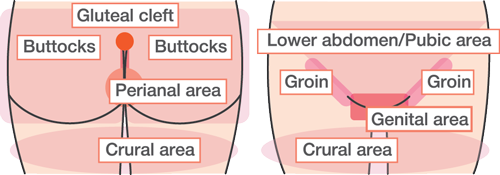
Figure 1. Common site of IAD (Created based on reference 1~3)
The structure and the role of the skin
The skin has a two-layer structure consisting of the epidermis and the dermis. The outer layer is mainly composed of keratinocytes, which mature, move towards the surface and eventually form corneocytes that make up the stratum corneum. In the stratum corneum, intercellular lipids fill the spaces between corneocytes, and the cells in the dermis are tightly bound together. This structure prevents excessive water loss and protects against the invasion of foreign substances, such as bacteria and irritants.
In this way, the epidermis plays an important role in the skin's barrier function.
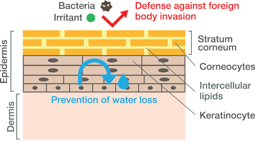
Figure 2. Structure and role of skin (Created based on reference 4)
Pathogenesis (Figure: 3)
・Disruption of skin barrier function due to skin maceration
If urine or feces (hereinafter referred to as excreta) touch the skin for a long time, the fluid of excreta causes the corneocytes to bulge, the intercellular lipids to decrease, and the chink of the corneocytes to expand. Furthermore, intercellular lipids are decomposed by fecal lipase (a lipid-degrading enzyme), and 'skin maceration' occurs. Macerated skin has a reduced barrier function, making it more susceptible to invasion by bacteria and irritants. It is also less resistant to friction; thus, the outer layer is easily damaged when it comes into contact with clothing, incontinence pads, and so on.
・Internal skin damage caused by the invasion of digestive enzymes and bacteria
The reduced barrier function of the skin, the digestive enzymes and the bacteria in feces invading the skin, can cause the following damages:
1. Tissue haemorrhage induced by digestive enzymes
The proteases in faeces (proteolytic enzymes) can penetrate the skin in which they degrade cells and the connections between them, leading to a further reduction in the skin's barrier function. Furthermore, the capillary walls are degraded, and bleeding occurs in the tissue. Mild rubor occurs without loss of the skin surface.
2. Bacterial damage to dermal tissue
Due to skin maceration and the reduction in skin barrier function caused by proteases, bacteria actively invade and proliferate in the skin, forming aggregated masses and damaging the underlying dermis. As a result, the dermis becomes fragile and can be easily disrupted, even by slight force, leading to the formation of blisters and erosion.
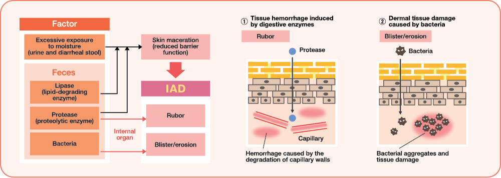
Figure 3: Pathogenesis of IAD (Created based on reference 4)
Multiple effects of excreta
Urea in urine is degraded into ammonia and carbon dioxide by urease (a urea-degrading enzyme) and digestive enzymes in faeces. Normally, the skin maintains a mildly acidic pH. However, ammonia, which is alkaline, increases the skin's pH. This promotes bacterial growth and increases the risk of IAD. An elevated pH also enhances the activity of digestive enzymes, further increasing the risk of skin damage. Furthermore, watery stools contain a large amount of digestive enzymes and therefore have a higher skin-damaging potential than hard stools.
Risk of accrual and assessment
The risk of IAD accrual is approximately 5.5 times higher in urine with a strong odour, while it is approximately 9.7 times higher in loose and watery stool. They have been reported to be more likely to cause IAD than when these factors are absent. Therefore, it is important to assess excreta to control/prevent IAD.
・Urine assessment
During urine assessment, we check for a strong odour (ammonia). Normal urine does not have a strong odour. However, when urine is infected with urease-producing bacteria, these bacteria produce ammonia, resulting in a characteristic strong smell. Therefore, the presence of a strong odour may indicate an increased risk of IAD.
・Faeces assessment
Loose and watery stools are associated with a high risk of IAD.
The Bristol Stool Form Scale is recommended for the objective and consistent assessment of stool conditions. This scale classifies stool into seven types (1-7), with types 5 and 6 indicating loose stool, and type 7 indicating watery stool.
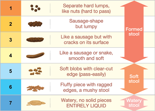
Figure 4: Bristol Stool Chart (Created based on reference 1,2)
Skincare for prevention and control
The standard approach to IAD prevention and management includes bed baths and cleansing (lavage). When formed stool or normal urine adheres to the skin, standard skincare should be provided. However, in cases of loose stool, watery stool, or strongly odorous urine, it is important to add a protective, water-repellent barrier to the standard skincare.
Bed-bath
- Cleansing should be performed after each episode of excretion (incontinence). Gently remove excreta using wet wipes or a skin cleanser.
- Avoid strong rubbing and do not use towels to reduce mechanical irritation to the skin.
- Use a skin cleanser when excreta cannot be easily removed with wet wipes. Wet wipes and skin cleansers containing oil are recommended, as they reduce friction and help minimise mechanical irritation to the skin.
- Rinsing with tepid water is recommended when excreta cannot be adequately removed by bed bathing alone.
Cleansing
- Excreta and scales should be cleaned once a day using a skin cleanser. ※Even when the frequency of urination and defecation is high, the use of skin cleansers should be limited to no more than once a day to prevent increased skin irritation.
- To prevent irritation to the skin, use a mildly acidic skin cleanser with a pH close to that of the skin (pH 5.5-7.0).
- Gently cleanse using the hands with form, and do not scrub strongly. Do not use a sponge or a nylon towel to reduce mechanical irritation to the skin.
- The skin cleanser should be washed off adequately with tepid water so that it will not remain on the skin.
- Dry the skin by gently patting, without rubbing, after washing.
Moisturising
- Apply a moisturiser more than once a day, especially after washing or bathing, as cleansing can remove natural skin oils.
- Use a moisturiser that contains emollient ingredients (e.g. petrolatum or mineral oil), which reduce transepidermal water loss and support intercellular lipids. Moisturisers containing ceramides or natural moisturising factors (NMF) are effective in enhancing and restoring skin barrier function.
- Moisturisers containing humectants (e.g. glycerine and urea), which attract water, should not be used on macerated skin.
- Apply moisturisers to all areas that may come into contact with excreta.
Protection (water-repellent)
- The frequency and timing should follow the manufacturer's instructions, while also taking into account the condition of the water-repellent barrier.
- Apply a water-repellent skin protectant containing petrolatum and dimethicone to prevent excreta from adhering to the skin.
- Use a skin barrier film containing acrylic copolymers when it is necessary to secure a dressing, such as in pressure ulcers, as adhesive products cannot be used over skin protectants. Skin protectants should be used in all areas where excreta may adhere.
-
Publication Date:August 04, 2026
-
Category:Skincare
HosCom International 2026 Vol. 2
